Stony Brook University Vibrational Spectroscopy Laboratory
Planetary Spectroscopy Group
 Welcome to the Planetary Spectroscopy Research Group and the Vibrational Spectroscopy Laboratory at Stony Brook University, led by Prof. Timothy Glotch. We have a variety of research interests with a focus using vibrational spectroscopy to understand the composition of Mars and the Moon.
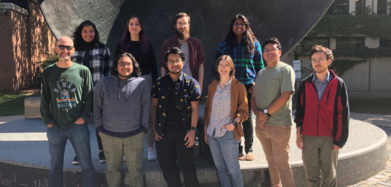
Back row, left to right: Nandita Kumari, Laura Breitenfeld, Connor Tinker, Indhu Varatharajan. Front row, left to right: Tim Glotch, Eashan Das, Anthony Munoz, Olive Koren, Leonard Flores, Chris Kremer.
Commitment to Diversity and Inclusion
The Center for Planetary Exploration and the Vibrational Spectroscopy Laboratory welcome students, postdoctoral researchers and visiting scholars regardless of race, religion, gender identification, sexual orientation, age, or disability status.
Group Research Interests
Remote Sensing of Mars, the Moon, and Small Bodies:
We use a variety of spacecraft and rover data sets to understand the crustal composition of Mars and the Moon as well as small bodies, including the near-Earth asteroid Bennu and Phobos, the largest moon of Mars. The mineralogy derived from these data sets provides insight into these bodies’ formation and alteration history.
Laboratory Spectroscopic Analysis of Martian, Lunar and Chondrite Samples and Analog Materials:
The spectroscopic tools available in the Vibrational Spectroscopy Laboratory allow us to examine geologic materials similar to those that are present on Mars, the Moon, and small solar system bodies. If we can understand the detailed spectral properties of these materials as well as what causes changes in these spectral properties, we can better interpret the remote sensing data.
Theoretical Calculations of Mineral Optical Constants and Scattering Properties:
We use several models to calculate the VNIR and MIR optical constants (n and k) of a variety of minerals of geologic interest. We are also using the publicly available Multiple Sphere T-Maxrix Model to exactly calculate the scattering properties of clusters of spheres of different compositions. Such studies enable the quantitative interpretation of remote sensing data from Earth, Mars, the Moon, and small bodies.
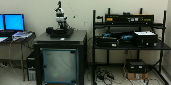
Vibrational Spectroscopy Laboratory Facilities
Neaspec NeaSNOM Nano-IR Imager and Spectrometer
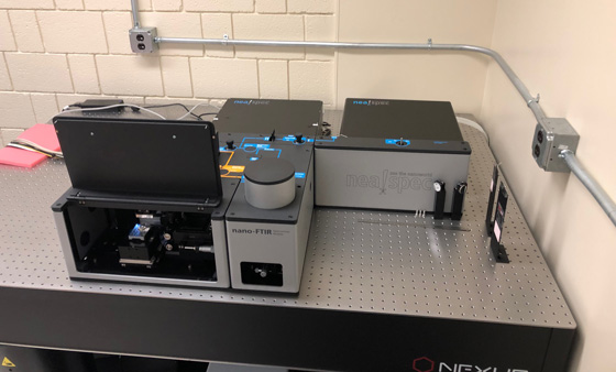
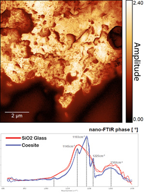 The neaspec NeaSNOM combines two infrared laser sources and an atomic force microscope (AFM) to enable broadband nano-IR imaging, spectroscopy, and hyperspectral imaging over the ~1.5-15 μm wavelength range at spatial scales of ~3-50 nm/pixel. The instrument simultaneously collects amplitude (reflectance) and phase (absorbance) data, maximizing the interpretability of the dataset. Spectral sampling of 3.3 cm-1 can be achieved across the entire spectral range, which is comparable to typical laboratory MIR far-field spectral sampling (~ 2 cm -1) for geological materials, superior to typical MIR emission spectrometers flown on planetary missions, and well within the range (<~20 cm-1 resolution) needed to resolve broad MIR bands. This spectral sampling also translates to ~1-2.5 nm sampling over the NIR range, which is also comparable to typical laboratory NIR reflectance measurements and superior to VNIR hyperspectral remote sensing data sets. The figure to the left shows a broadband nano-IR amplitude image of a portion of a shocked rock from Lonar crater, India and characteristic spectra from the bright (coesite) and dark (glass) regions, demonstrating substantial shock heterogeneity at sub-micron spatial scales.
Nicolet 6700 FTIR Spectrometer

 The Thermo Fisher Nicolet 6700 FTIR spectrometer is modified to collect emissivity spectra in an environment purged of water vapor and CO2. Measurements from 400-4000 cm-1 can be carried out using a KBr beamsplitter and a deuterated L-alanine doped triglycine sulfate (DLaTGS) detector with a KBr window. Measurements from 50-600 cm-1 can be carried out on the same spectrometer equipped with a Solid Substrate beamsplitter and a DTGS detector with a polyethylene window. The spectrometer can be equipped with a SmartOrbit attenuated total reflectance (ATR) accessory with a Type IIa diamond element for ATR analyses or a FT-30 accessory for specular reflectance measurements. The spectrometer can also be equipped with a CaF2 beamsplitter, an InGaAS detector, and a Smart Diffuse Reflectance accessory for diffuse reflectance measurements from 0.8-2.5 μm.
Nicolet iN10MX Micro-Imaging FTIR Spectrometer

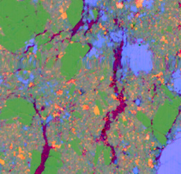
The Nicolet iN10MX FTIR microscope is equipped with a liquid nitrogen-cooled MCT array detector capable of acquiring hyperspectral image cubes between 7000 and 715 cm-1. Pixel sizes are 25 or 6 μm for reflectance and transmission modes and 25 or 1.3 μm for ATR mode. The iN10MX is also equipped with a DTGS detector capable of acquiring 50 x 50 μm or larger point spectra between 4000 and 400 cm-1. The image above is a 2 mm by 2 mm reflectance spectral image of a shocked basalt from Lonar Crater, India. Green pixels are maskelynite, light blue pixels are melt glass, orange pixels are pyroxene, and purple pixels are epoxy.
WITec alpha300R Micro-Imaging Raman Spectrometer
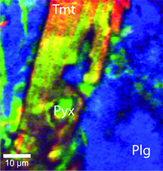 The WITec alpha300R confocal Raman imaging system is equipped with both 532 nm Nd YAG and 785 nm diode lasers. A variety objective lenses provide white light and Raman imaging capability at scales ranging from 260 nm/pixel to several μm/pixel. Hyperspectral Raman maps or point spectra can be acquired on mineral or biological samples as well as high pressure materials within diamond anvil cells. Two holographic grating spectrometers allow the collection of Raman spectra at 3 cm-1 or 1 cm-1 spectral sampling over a wide Δcm-1 range. The image to the left is a high-resolution Raman map of a flood basalt sample from Lonar, India, showing the distributions of titanomagnetite, plagioclase feldspar, and pyroxene.